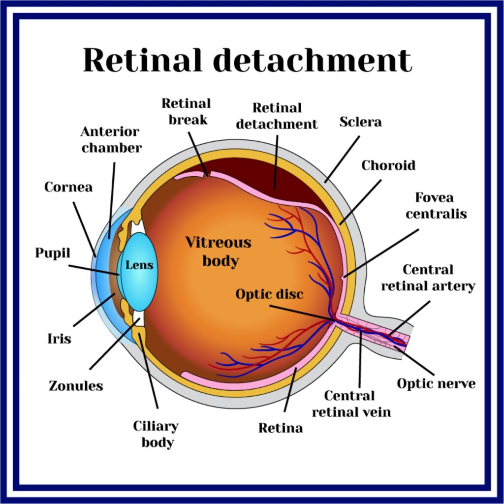
Retinal detachment
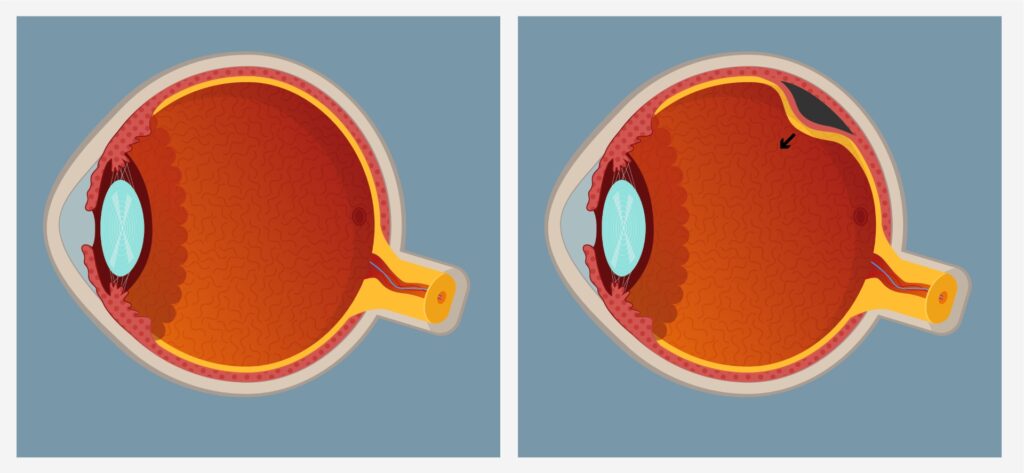
In patients who have a retinal crack, fluid from the vitreous cavity may move through the rupture below the retina, causing retinal detachment. This slide shows a detachment that shows folds of the retina and a loss of some transparency of the inner retinal tissue so that the underlying orange color is less visible.
Patients with retinal detachment often observe a killing in the part of their vision that is affected by detachment flashes (photopsies) or myopias (flies). Retinal detachments are progressive, initially affecting only a portion of vision but over time they will affect more. If the macula is detached, the central visual acuity will be lost and there is an increased risk of permanent vision loss.
Retinal fissure can be treated with Laser retinal detachment (incision crack with photocoagulation), while when a concomitant retinal detachment has been induced, it is treated depending on the case with some surgical technique from the following: (1) cavity, or (2) removal of the vitreous (vitrectomy) by laser surgery or cryocoagulation applied to areas with a retinal fissure; (3) suturing of a silicone band around the eye to support the retina (scle).


